

2mo
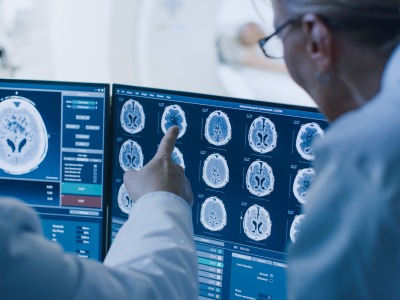
Did you know? Precision neurology is transforming care by integrating genomics, biomarkers, advanced imaging, and computational modeling to tailor diagnosis and treatment at the individual level. By moving beyond population averages, this approach enables earlier detection, better trial stratification, and more personalized therapeutic decisions across conditions like Alzheimer’s, Parkinson’s, epilepsy, and stroke.
How might integrating multi-omics and brain imaging reshape personalized decision-making in everyday neurological practice?
How might integrating multi-omics and brain imaging reshape personalized decision-making in everyday neurological practice?